
Featured publications
Opioid- and NMDA-receptor-dependent neural plasticity mediates long-term analgesia from motor cortical stimulation. Lindsay NM, Haziza S, Mackey S, Baer TM, Scherrer G, Schnitzer MJ. bioRxiv [Preprint]. 2026 Jul 7:2026.07.01.735554. PMID: 42465307. DOI: 10.64898/2026.07.01.735554.
Human assembloid model of the ascending neural sensory pathway. Kim J, Imaizumi K, Jurjuț O, Kelley KW, Wang D, Thete M, Hudacova Z, Amin ND, Levy RJ, Scherrer G, & Pașca S. Nature. 2025 Apr 9. PMID: 40205039. DOI: 10.1038/s41586-025-08808-3.
Neural circuit basis of placebo pain relief. Chen C, Niehaus JK, Dinc F, Huang KL, Barnette AL, Tassou A, Shuster SA, Wang L, Lemire A, Menon V, Ritola K, Hantman AW, Zeng H, Schnitzer MJ, Scherrer G. Nature. 2024 Jul 24. DOI: 10.1038/s41586-024-07816-z. PMID: 39048016.
Scientific Impact: Harnessing expectations to relieve pain. We discovered a previously unknown brain circuit through which the expectation of pain relief engages the brain’s own pain-control systems. Positive expectations activate a pathway connecting the anterior cingulate cortex to the pons and cerebellum, ultimately reducing pain. These findings provide a biological mechanism for how beliefs and positive expectations can influence the experience of pain and suggest a neural substrate through which cognitive approaches such as cognitive behavioral therapy (CBT) may produce pain relief. We are now developing pharmacological and neuromodulatory approaches to engage this pathway and leverage the brain’s endogenous analgesic systems to treat acute post-operative pain and chronic pain.
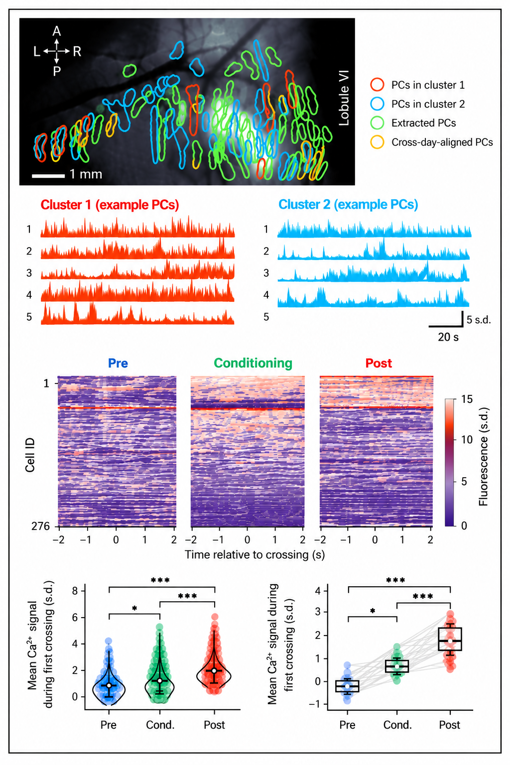
Figure: Purkinje cells encode expectations of pain relief. Calcium imaging revealed that Purkinje cells in cognitive regions of the cerebellum, which receive input from the cingulate cortex through the pons, become strongly engaged during learning that a specific action leads to pain reduction. Their activity changes as this association is learned, revealing how the expectation of pain relief is represented at the level of individual cerebellar neurons.
Media articles and podcasts. This study has attracted exceptional international attention, ranking in the top 1% of all articles of a similar age and the top 6% of articles published in Nature.
Structure-guided design of a peripherally restricted chemogenetic system. Kang HJ, Krumm BE, Tassou A, Geron M, DiBerto JF, Kapolka NJ, Gumpper RH, Sakamoto K, Kocak DD, Olsen RHJ, Huang XP, Zhang S, Huang KL, Zaidi SA, Nguyen MTV, Jo MJ, Katritch V, Fay JF, Scherrer G, Roth BL. Cell. 2024 Dec 26;187(26):7433-7449.e20. PMID: 39631393. PMCID: PMC12291529. DOI: 10.1016/j.cell.2024.11.001
Scientific Impact: Toward precision control of pain-sensing neurons. In collaboration with Bryan Roth’s laboratory (UNC-CH), we helped develop HCAD, a new chemogenetic system for precisely controlling peripheral cells. Our laboratory developed viral vectors with specific genetic elements to optimize HCAD expression in dorsal root ganglion neurons and performed the anatomical, electrophysiological, and behavioral studies demonstrating its ability to inhibit pain-sensing neurons in vivo. This technology could ultimately allow us to selectively silence the specific classes of sensory neurons that drive a chronic pain condition rather than broadly suppressing pain sensation throughout the body. The goal is to relieve ongoing pathological pain while preserving the protective pain signals that warn of a new injury or disease. More broadly, DREADD-based chemogenetics is now beginning to move toward human translation, opening the possibility of using genetically targeted receptors to control disease-relevant cells with unprecedented precision.
Hyperexcitable arousal circuits drive sleep instability during aging. Li SB, Damonte VM, Chen C, Wang GX, Kebschull JM, Yamaguchi H, Bian WJ, Purmann C, Pattni R, Urban AE, Mourrain P, Kauer JA, Scherrer G, de Lecea L. Science. 2022 Feb 25;375(6583). doi: 10.1126/science.abh3021. PMID: 35201886
Brain circuits for pain and its treatment. Mercer Lindsay N, Chen C, Gilam G, Mackey S, Scherrer G. Science Translational Medicine. 2021 Nov 10;13(619). doi: 10.1126/scitranslmed.abj7360. PMID: 34757810
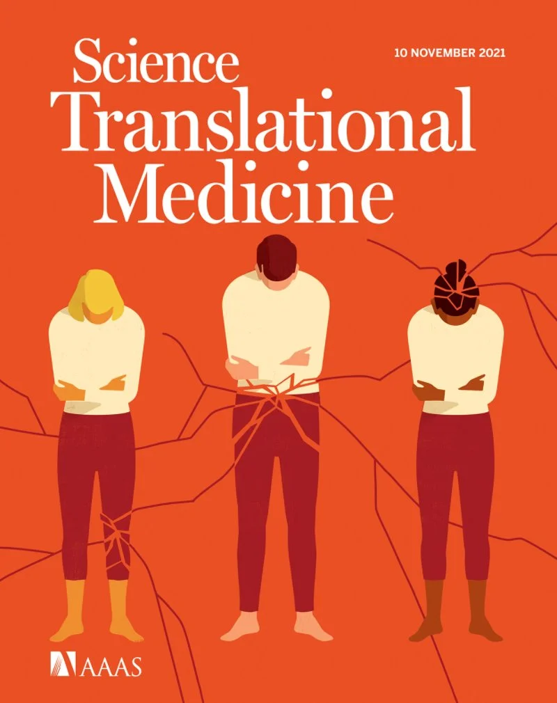
On the cover. Pain, Pain Go Away. The cover image symbolizes the shattering experience of different types of pain in the human body. In this Special Issue, which is designed to showcase the latest advances in pain research, Review articles discuss the principal brain circuits mediating pain (Lindsay et al.) and insights and challenges associated with the development of analgesic drugs (Jayakar et al.). In addition, three Viewpoint articles highlight promising areas of pain research including inflammatory mechanisms of pain (Kavelaars et al.), sexual dimorphism in pain sensitivity (Navratilova et al.), and development of imaging-based biomarkers for measuring pain (Tracey). Two Research Articles complete the Special Issue, presenting research findings on a new small molecule with analgesic effects in mouse models of pain (Cai et al.) and a cytokine that contributes to inflammatory itch (Tseng and Hoon). These Special Issue articles showcase the encouraging progress made in the field of pain research and the challenges still to be overcome. Credit: Chris Gash.
An amygdalar neural ensemble that encodes the unpleasantness of pain. Corder G*, Ahanonu B*, Grewe BF, Wang D, Schnitzer MJ, Scherrer G. Science. 2019 Jan 18;363(6424):276-281. *Co-first authors. doi: 10.1126/science.aap8586. PMID: 30655440
Scientific Impact: Separating pain from suffering. We discovered a population of neurons in the amygdala—a brain region central to emotion—that encodes the unpleasantness of pain. Silencing these neurons reduced pain-related avoidance behaviors while preserving reflexes and the ability to detect potentially harmful stimuli. This discovery revealed that the suffering caused by pain can be targeted independently from the sensory warning signals that protect us from injury. We are now funded by the NIH HEAL Initiative to identify druggable receptors in these neurons and develop a new generation of non-addictive pain medicines designed to reduce suffering across different types of acute postoperative and chronic pain while preserving protective pain sensation.
Media articles and podcasts

Endogenous and exogenous opioids in pain. Corder G, Castro DC, Bruchas MR, Scherrer G. Annu Rev Neurosci. 2018 Jul 8;41:453-473. doi: 10.1146/annurev-neuro-080317-061522. Epub 2018 May 31. PMID: 29852083
Functional divergence of delta and mu opioid receptor organization in CNS pain circuits. Wang D, Tawfik VL, Corder G, Low SA, François A, Basbaum AI, Scherrer G. Neuron. 2018 Apr 4;98(1):90-108.e5. doi: 10.1016/j.neuron.2018.03.002. Epub 2018 Mar 22. PMID: 29576387
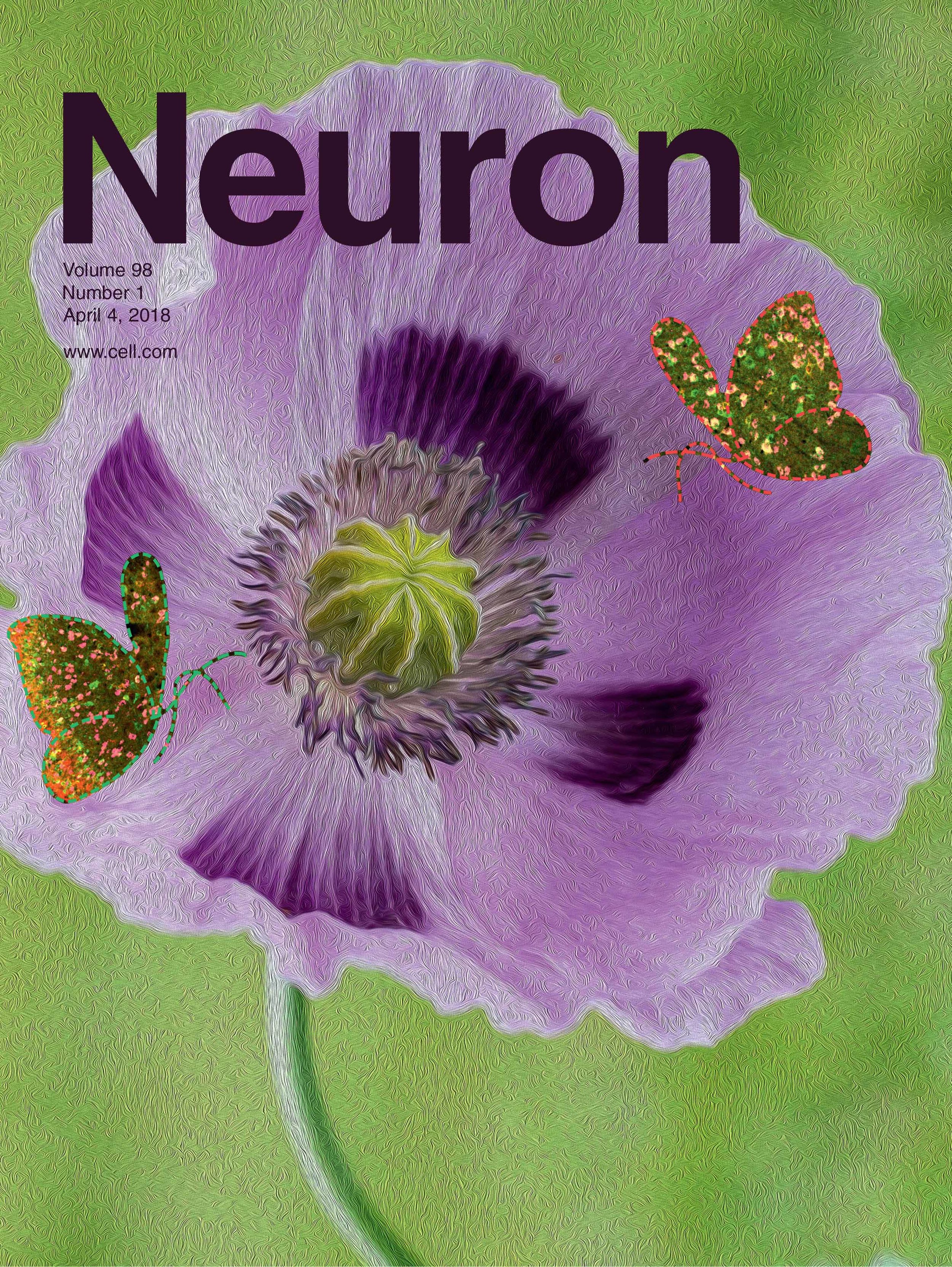
On the cover. Cellular interactions between delta and mu opioid receptors (DORs and MORs) are thought to regulate opioid analgesia. However, the neurons in which such interactions could occur within pain neural circuits in vivo remains elusive. In this issue of Neuron, Wang et al. showed that DOR and MOR are present in mostly different populations of nociceptive neurons in the spinal cord and brain, indicating that they may control distinct aspects of pain experience. The cover image illustrates the interactions between two butterflies, DOR and MOR, flying over an opium poppy flower. The butterflies’ wing color patterns show DOR-MOR segregated expression in dorsal horn pain neurons (left) and co-expression in ventral horn neurons constituting motor neural circuits (right). Artwork by Dong Wang.
Media articles
A brainstem-spinal cord inhibitory circuit for mechanical pain modulation by GABA and enkephalins. François A, Low SA, Sypek EI, Christensen AJ, Sotoudeh C, Beier KT, Ramakrishnan C, Ritola KD, Sharif-Naeini R, Deisseroth K, Delp SL, Malenka RC, Luo L, Hantman AW, Scherrer G. Neuron. 2017 Feb 22;93(4):822-839.e6. doi: 10.1016/j.neuron.2017.01.008. Epub 2017 Feb 2. PMID: 28162807
Loss of μ-opioid receptor signaling in nociceptors, but not microglia, abrogates morphine tolerance without disrupting analgesia. Corder G*, Tawfik VL*, Wang D*, Sypek EI*, Low SA, Dickinson JR, Sotoudeh C, Clark JD, Barres BA, Bohlen CJ, Scherrer G. Nature Medicine. 2017 Feb;23(2):164-173. *Co-first authors. doi: 10.1038/nm.4262. Epub 2017 Jan 16. PMID: 28092666
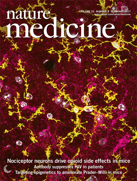
On the cover. Corder, Tawfik, Wang, Sypek et al. establish that mu opioid receptors (MORs) on peripheral nociceptor neurons, but not microglia, are responsible for opioid-related side-effects of tolerance and opioid-induced hyperalgesia. They further demonstrate that a commercially available peripherally-restricted opioid antagonist, methylnaltrexone bromide, mitigates these side effects when co-administered with morphine. Image depicts mouse spinal cord with Oprm1 mRNA (coding for MOR, fuchsia) concentrated in neurons but not microglia (CD11b, yellow). Image credit: Vivianne Tawfik.
Media articles
In vivo interrogation of spinal mechanosensory circuits. Christensen AJ, Iyer SM, François A, Vyas S, Ramakrishnan C, Vesuna S, Deisseroth K, Scherrer G, Delp SL. Cell Reports. 2016 Nov 1;17(6):1699-1710. doi: 10.1016/j.celrep.2016.10.010. PMID: 27806306
Structure-based discovery of opioid analgesics with reduced side effects. Manglik A, Lin H, Aryal DK, McCorvy JD, Dengler D, Corder G, Levit A, Kling RC, Bernat V, Hübner H, Huang XP, Sassano MF, Giguère PM, Löber S, Da Duan, Scherrer G, Kobilka BK, Gmeiner P, Roth BL, Shoichet BK. Nature. 2016 Sep 8;537(7619):185-190. doi: 10.1038/nature19112. Epub 2016 Aug 17. PMID: 27533032
Delta opioid receptors presynaptically regulate cutaneous mechanosensory neuron input to the spinal cord dorsal horn. Bardoni R, Tawfik VL, Wang D, François A, Solorzano C, Shuster SA, Choudhury P, Betelli C, Cassidy C, Smith K, de Nooij JC, Mennicken F, O'Donnell D, Kieffer BL, Woodbury CJ, Basbaum AI, MacDermott AB, Scherrer G. Neuron. 2014 Mar 19;81(6):1312-1327. doi: 10.1016/j.neuron.2014.01.044. Epub 2014 Feb 27. PMID: 24583022
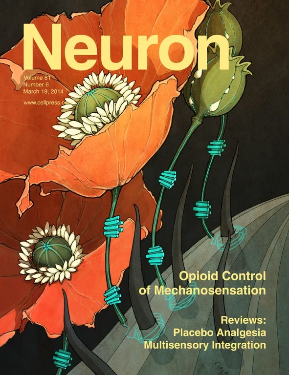
On the cover. Morphine and other mu opioid receptor agonists are gold standard analgesics due to their potent inhibitory action on pain processing mechanisms. In this issue, Bardoni et al. provide evidence that the opioid system also exercises broad control over cutaneous mechanosensation, via delta opioid receptors (DORs). The authors show that DOR, depicted in light blue, is expressed by mechanosensory neurons that constitute touch-encoding organs in the skin, including circumferential endings around hair follicles (dark gray). In these neurons, DORs inhibit voltage-gated calcium channels and dampen mechanosensory input to the central nervous system and could represent promising drug targets to combat touch-evoked neuropathic pain.
Media articles
Pre- and postsynaptic inhibitory control in the spinal cord dorsal horn. Bardoni R, Takazawa T, Tong CK, Choudhury P, Scherrer G, Macdermott AB. Ann N Y Acad Sci. 2013 Mar;1279:90-6. doi: 10.1111/nyas.12056. Review. PMID: 23531006
VGLUT2 expression in primary afferent neurons is essential for normal acute pain and injury-induced heat hypersensitivity. Scherrer G, Low SA, Wang X, Zhang J, Yamanaka H, Urban R, Solorzano C, Harper B, Hnasko TS, Edwards RH, Basbaum AI. Proc Natl Acad Sci U S A. 2010 Dec 21;107(51):22296-301. doi: 10.1073/pnas.1013413108. Epub 2010 Dec 6. PMID: 21135246
Cellular and molecular mechanisms of pain. Basbaum AI, Bautista DM, Scherrer G, Julius D. Cell. 2009 Oct 16;139(2):267-84. doi: 10.1016/j.cell.2009.09.028. Review. PMID: 19837031
Dissociation of the opioid receptor mechanisms that control mechanical and heat pain. Scherrer G, Imamachi N, Cao YQ, Contet C, Mennicken F, O'Donnell D, Kieffer BL, Basbaum AI. Cell. 2009 Jun 12;137(6):1148-59. doi: 10.1016/j.cell.2009.04.019. PMID: 19524516
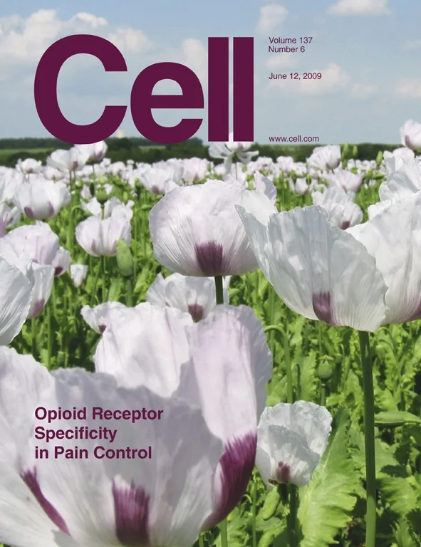
On the cover. How different opioid receptor subtypes regulate pain is unclear. It is generally considered that the delta and mu opioid receptors (DOR and MOR, respectively) co-occur in subsets of pain fibers (nociceptors), where they coordinately regulate pain. In this issue of Cell, Scherrer et al. demonstrate that the MOR and DOR are, in fact, expressed by different nociceptor populations. The authors further show that the segregated MOR and DOR distribution is paralleled by a selective functional contribution of the two receptors to the control of heat and mechanical pain, respectively. Photo credit: Opium poppy field in France, by Wolfgang Horlacher. Used under the terms of the GNU Free Documentation License.
Knockin mice expressing fluorescent delta-opioid receptors uncover G protein-coupled receptor dynamics in vivo. Scherrer G, Tryoen-Tóth P, Filliol D, Matifas A, Laustriat D, Cao YQ, Basbaum AI, Dierich A, Vonesh JL, Gavériaux-Ruff C, Kieffer BL. Proc Natl Acad Sci U S A. 2006 Jun 20;103(25):9691-6. Epub 2006 Jun 9. PMID: 16766653